Praseodymium »
PDB 1k0z-6b41 »
3ate »
Praseodymium in PDB 3ate: Crystal Structure of the KIR3.2 Cytoplasmic Domain (Na+-Free Crystal Soaked in 10 Mm Praseodymium (III) Acetate)
Protein crystallography data
The structure of Crystal Structure of the KIR3.2 Cytoplasmic Domain (Na+-Free Crystal Soaked in 10 Mm Praseodymium (III) Acetate), PDB code: 3ate
was solved by
A.Inanobe,
Y.Kurachi,
with X-Ray Crystallography technique. A brief refinement statistics is given in the table below:
| Resolution Low / High (Å) | 48.47 / 3.20 |
| Space group | I 4 2 2 |
| Cell size a, b, c (Å), α, β, γ (°) | 82.775, 82.775, 172.894, 90.00, 90.00, 90.00 |
| R / Rfree (%) | 26.7 / 32.5 |
Praseodymium Binding Sites:
The binding sites of Praseodymium atom in the Crystal Structure of the KIR3.2 Cytoplasmic Domain (Na+-Free Crystal Soaked in 10 Mm Praseodymium (III) Acetate)
(pdb code 3ate). This binding sites where shown within
5.0 Angstroms radius around Praseodymium atom.
In total 3 binding sites of Praseodymium where determined in the Crystal Structure of the KIR3.2 Cytoplasmic Domain (Na+-Free Crystal Soaked in 10 Mm Praseodymium (III) Acetate), PDB code: 3ate:
Jump to Praseodymium binding site number: 1; 2; 3;
In total 3 binding sites of Praseodymium where determined in the Crystal Structure of the KIR3.2 Cytoplasmic Domain (Na+-Free Crystal Soaked in 10 Mm Praseodymium (III) Acetate), PDB code: 3ate:
Jump to Praseodymium binding site number: 1; 2; 3;
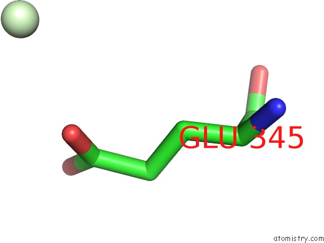
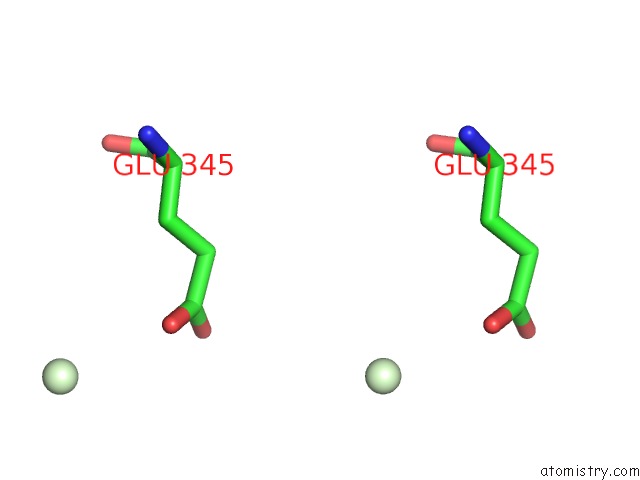
Praseodymium binding site 1 out of 3 in 3ate
Go back to
Praseodymium binding site 1 out
of 3 in the Crystal Structure of the KIR3.2 Cytoplasmic Domain (Na+-Free Crystal Soaked in 10 Mm Praseodymium (III) Acetate)
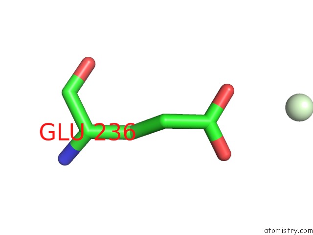
Mono view
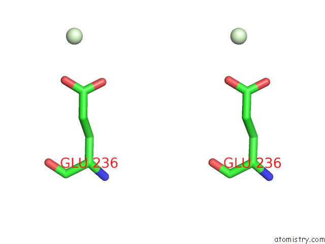
Stereo pair view
Mono view
Stereo pair view
A full contact list of Praseodymium with other atoms in the Pr binding
site number 1 of Crystal Structure of the KIR3.2 Cytoplasmic Domain (Na+-Free Crystal Soaked in 10 Mm Praseodymium (III) Acetate) within 5.0Å range:
|
Praseodymium binding site 2 out of 3 in 3ate
Go back to
Praseodymium binding site 2 out
of 3 in the Crystal Structure of the KIR3.2 Cytoplasmic Domain (Na+-Free Crystal Soaked in 10 Mm Praseodymium (III) Acetate)
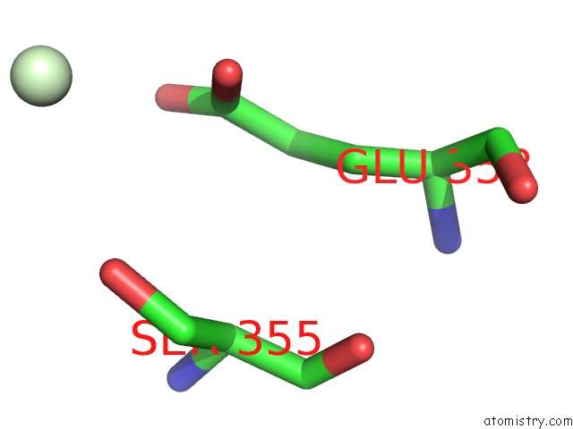
Mono view
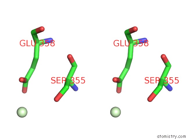
Stereo pair view
Mono view
Stereo pair view
A full contact list of Praseodymium with other atoms in the Pr binding
site number 2 of Crystal Structure of the KIR3.2 Cytoplasmic Domain (Na+-Free Crystal Soaked in 10 Mm Praseodymium (III) Acetate) within 5.0Å range:
|
Praseodymium binding site 3 out of 3 in 3ate
Go back to
Praseodymium binding site 3 out
of 3 in the Crystal Structure of the KIR3.2 Cytoplasmic Domain (Na+-Free Crystal Soaked in 10 Mm Praseodymium (III) Acetate)
Mono view
Stereo pair view
Mono view
Stereo pair view
A full contact list of Praseodymium with other atoms in the Pr binding
site number 3 of Crystal Structure of the KIR3.2 Cytoplasmic Domain (Na+-Free Crystal Soaked in 10 Mm Praseodymium (III) Acetate) within 5.0Å range:
|
Reference:
A.Inanobe,
A.Nakagawa,
Y.Kurachi.
Interactions of Cations with the Cytoplasmic Pores of Inward Rectifier K(+) Channels in the Closed State J.Biol.Chem. V. 286 41801 2011.
ISSN: ISSN 0021-9258
PubMed: 21982822
DOI: 10.1074/JBC.M111.278531
Page generated: Thu Oct 10 10:25:31 2024
ISSN: ISSN 0021-9258
PubMed: 21982822
DOI: 10.1074/JBC.M111.278531
Last articles
Zn in 9MJ5Zn in 9HNW
Zn in 9G0L
Zn in 9FNE
Zn in 9DZN
Zn in 9E0I
Zn in 9D32
Zn in 9DAK
Zn in 8ZXC
Zn in 8ZUF