Praseodymium »
PDB 1k0z-6b41 »
3u3u »
Praseodymium in PDB 3u3u: Crystal Structure of the Tablysin-15-Leukotriene E4 Complex
Protein crystallography data
The structure of Crystal Structure of the Tablysin-15-Leukotriene E4 Complex, PDB code: 3u3u
was solved by
J.F.Andersen,
with X-Ray Crystallography technique. A brief refinement statistics is given in the table below:
| Resolution Low / High (Å) | 17.45 / 2.50 |
| Space group | P 31 2 1 |
| Cell size a, b, c (Å), α, β, γ (°) | 69.774, 69.774, 85.473, 90.00, 90.00, 120.00 |
| R / Rfree (%) | 20.2 / 25.3 |
Praseodymium Binding Sites:
The binding sites of Praseodymium atom in the Crystal Structure of the Tablysin-15-Leukotriene E4 Complex
(pdb code 3u3u). This binding sites where shown within
5.0 Angstroms radius around Praseodymium atom.
In total only one binding site of Praseodymium was determined in the Crystal Structure of the Tablysin-15-Leukotriene E4 Complex, PDB code: 3u3u:
In total only one binding site of Praseodymium was determined in the Crystal Structure of the Tablysin-15-Leukotriene E4 Complex, PDB code: 3u3u:
Praseodymium binding site 1 out of 1 in 3u3u
Go back to
Praseodymium binding site 1 out
of 1 in the Crystal Structure of the Tablysin-15-Leukotriene E4 Complex
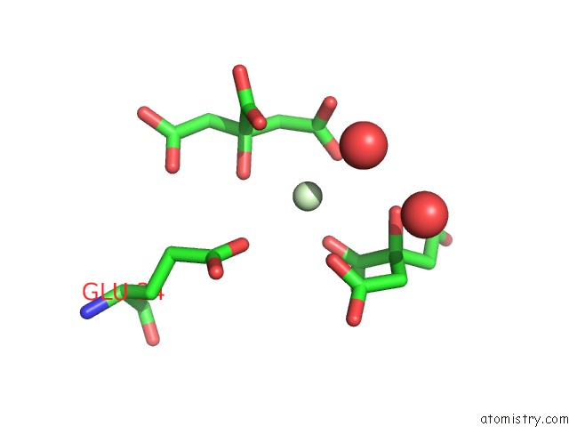
Mono view
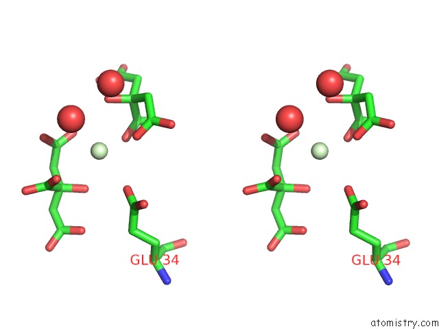
Stereo pair view
Mono view
Stereo pair view
A full contact list of Praseodymium with other atoms in the Pr binding
site number 1 of Crystal Structure of the Tablysin-15-Leukotriene E4 Complex within 5.0Å range:
|
Reference:
X.Xu,
I.M.Francischetti,
R.Lai,
J.M.Ribeiro,
J.F.Andersen.
Structure of Protein Having Inhibitory Disintegrin and Leukotriene Scavenging Functions Contained in Single Domain. J.Biol.Chem. V. 287 10967 2012.
ISSN: ISSN 0021-9258
PubMed: 22311975
DOI: 10.1074/JBC.M112.340471
Page generated: Thu Oct 10 10:25:59 2024
ISSN: ISSN 0021-9258
PubMed: 22311975
DOI: 10.1074/JBC.M112.340471
Last articles
Zn in 9MJ5Zn in 9HNW
Zn in 9G0L
Zn in 9FNE
Zn in 9DZN
Zn in 9E0I
Zn in 9D32
Zn in 9DAK
Zn in 8ZXC
Zn in 8ZUF