Praseodymium »
PDB 1k0z-6b41 »
2f1r »
Praseodymium in PDB 2f1r: Crystal Structure of Molybdopterin-Guanine Biosynthesis Protein B (Mobb)
Protein crystallography data
The structure of Crystal Structure of Molybdopterin-Guanine Biosynthesis Protein B (Mobb), PDB code: 2f1r
was solved by
L.Damodharan,
S.Eswaramoorthy,
D.Kumaran,
S.Swaminathan,
S.K.Burley,
New York Sgx Research Center For Structuralgenomics (Nysgxrc),
with X-Ray Crystallography technique. A brief refinement statistics is given in the table below:
| Resolution Low / High (Å) | 50.00 / 2.10 |
| Space group | P 1 21 1 |
| Cell size a, b, c (Å), α, β, γ (°) | 37.980, 63.930, 66.010, 90.00, 95.30, 90.00 |
| R / Rfree (%) | 18.6 / 25.3 |
Other elements in 2f1r:
The structure of Crystal Structure of Molybdopterin-Guanine Biosynthesis Protein B (Mobb) also contains other interesting chemical elements:
| Chlorine | (Cl) | 1 atom |
Praseodymium Binding Sites:
The binding sites of Praseodymium atom in the Crystal Structure of Molybdopterin-Guanine Biosynthesis Protein B (Mobb)
(pdb code 2f1r). This binding sites where shown within
5.0 Angstroms radius around Praseodymium atom.
In total only one binding site of Praseodymium was determined in the Crystal Structure of Molybdopterin-Guanine Biosynthesis Protein B (Mobb), PDB code: 2f1r:
In total only one binding site of Praseodymium was determined in the Crystal Structure of Molybdopterin-Guanine Biosynthesis Protein B (Mobb), PDB code: 2f1r:
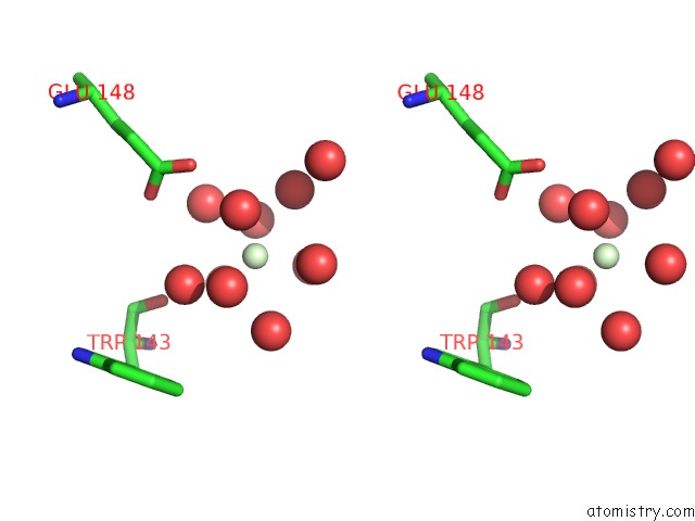
Praseodymium binding site 1 out of 1 in 2f1r
Go back to
Praseodymium binding site 1 out
of 1 in the Crystal Structure of Molybdopterin-Guanine Biosynthesis Protein B (Mobb)
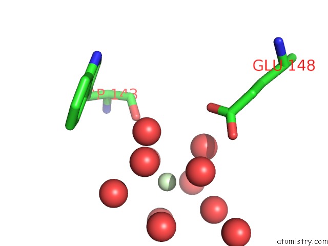
Mono view
Stereo pair view
Mono view
Stereo pair view
A full contact list of Praseodymium with other atoms in the Pr binding
site number 1 of Crystal Structure of Molybdopterin-Guanine Biosynthesis Protein B (Mobb) within 5.0Å range:
|
Reference:
L.Damodharan,
S.Eswaramoorthy,
D.Kumaran,
S.Swaminathan.
Crystal Structure of Molybdopterin-Guanine Dinucleotide Biosynthesis Protein B (Mobb) To Be Published.
Page generated: Thu Oct 10 10:24:37 2024
Last articles
K in 8QXTK in 8QXS
K in 8QUD
K in 8QUC
K in 8QRN
K in 8QRK
K in 8QRM
K in 8QRL
K in 8QOS
K in 8QN2