Praseodymium »
PDB 1k0z-6b41 »
2wvi »
Praseodymium in PDB 2wvi: Crystal Structure of the N-Terminal Domain of BUBR1
Enzymatic activity of Crystal Structure of the N-Terminal Domain of BUBR1
All present enzymatic activity of Crystal Structure of the N-Terminal Domain of BUBR1:
2.7.11.1;
2.7.11.1;
Protein crystallography data
The structure of Crystal Structure of the N-Terminal Domain of BUBR1, PDB code: 2wvi
was solved by
S.D'arcy,
O.R.Davies,
T.L.Blundell,
V.M.Bolanos-Garcia,
with X-Ray Crystallography technique. A brief refinement statistics is given in the table below:
| Resolution Low / High (Å) | 54.39 / 1.80 |
| Space group | P 32 2 1 |
| Cell size a, b, c (Å), α, β, γ (°) | 62.796, 62.796, 90.450, 90.00, 90.00, 120.00 |
| R / Rfree (%) | 22.188 / 25.986 |
Praseodymium Binding Sites:
The binding sites of Praseodymium atom in the Crystal Structure of the N-Terminal Domain of BUBR1
(pdb code 2wvi). This binding sites where shown within
5.0 Angstroms radius around Praseodymium atom.
In total 2 binding sites of Praseodymium where determined in the Crystal Structure of the N-Terminal Domain of BUBR1, PDB code: 2wvi:
Jump to Praseodymium binding site number: 1; 2;
In total 2 binding sites of Praseodymium where determined in the Crystal Structure of the N-Terminal Domain of BUBR1, PDB code: 2wvi:
Jump to Praseodymium binding site number: 1; 2;
Praseodymium binding site 1 out of 2 in 2wvi
Go back to
Praseodymium binding site 1 out
of 2 in the Crystal Structure of the N-Terminal Domain of BUBR1
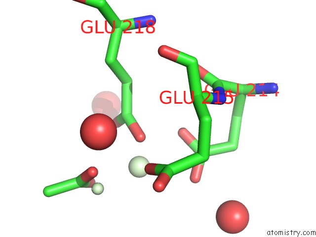
Mono view
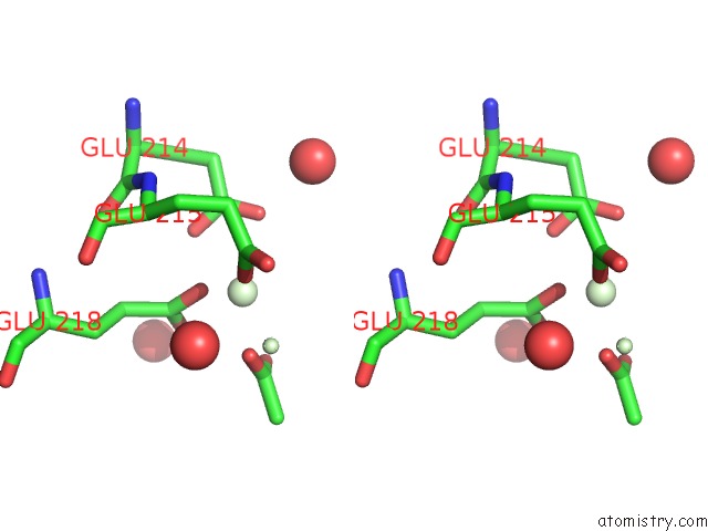
Stereo pair view
Mono view
Stereo pair view
A full contact list of Praseodymium with other atoms in the Pr binding
site number 1 of Crystal Structure of the N-Terminal Domain of BUBR1 within 5.0Å range:
|
Praseodymium binding site 2 out of 2 in 2wvi
Go back to
Praseodymium binding site 2 out
of 2 in the Crystal Structure of the N-Terminal Domain of BUBR1
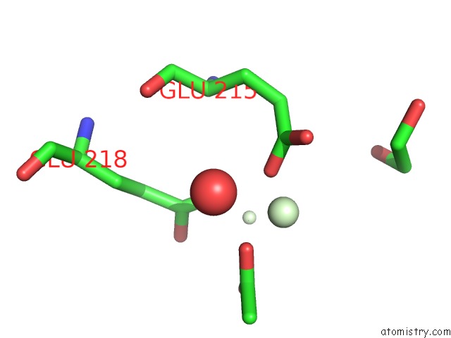
Mono view
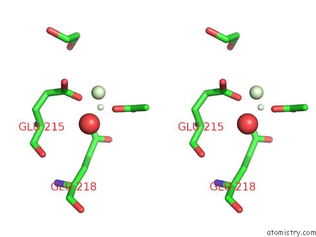
Stereo pair view
Mono view
Stereo pair view
A full contact list of Praseodymium with other atoms in the Pr binding
site number 2 of Crystal Structure of the N-Terminal Domain of BUBR1 within 5.0Å range:
|
Reference:
S.D'arcy,
O.R.Davies,
T.L.Blundell,
V.M.Bolanos-Garcia.
Defining the Molecular Basis of BUBR1 Kinetochore Interactions and Anaphase-Promoting Complex/Cyclosome (Apc/C)-CDC20 Inhibition J.Biol.Chem. V. 285 14764 2010.
ISSN: ISSN 0021-9258
PubMed: 20220147
DOI: 10.1074/JBC.M109.082016
Page generated: Thu Oct 10 10:24:37 2024
ISSN: ISSN 0021-9258
PubMed: 20220147
DOI: 10.1074/JBC.M109.082016
Last articles
Mn in 7L3VMn in 7LAF
Mn in 7L3M
Mn in 7L6R
Mn in 7L36
Mn in 7KTM
Mn in 7L29
Mn in 7L28
Mn in 7KTL
Mn in 7L27