Praseodymium »
PDB 6e1a-9bif »
6oop »
Praseodymium in PDB 6oop: Protein B
Protein crystallography data
The structure of Protein B, PDB code: 6oop
was solved by
M.Lu,
with X-Ray Crystallography technique. A brief refinement statistics is given in the table below:
| Resolution Low / High (Å) | 15.00 / 2.80 |
| Space group | C 1 2 1 |
| Cell size a, b, c (Å), α, β, γ (°) | 95.068, 63.149, 102.074, 90.00, 100.87, 90.00 |
| R / Rfree (%) | n/a / n/a |
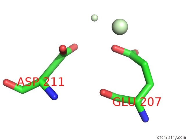
Praseodymium Binding Sites:
The binding sites of Praseodymium atom in the Protein B
(pdb code 6oop). This binding sites where shown within
5.0 Angstroms radius around Praseodymium atom.
In total 2 binding sites of Praseodymium where determined in the Protein B, PDB code: 6oop:
Jump to Praseodymium binding site number: 1; 2;
In total 2 binding sites of Praseodymium where determined in the Protein B, PDB code: 6oop:
Jump to Praseodymium binding site number: 1; 2;
Praseodymium binding site 1 out of 2 in 6oop
Go back to
Praseodymium binding site 1 out
of 2 in the Protein B

Mono view
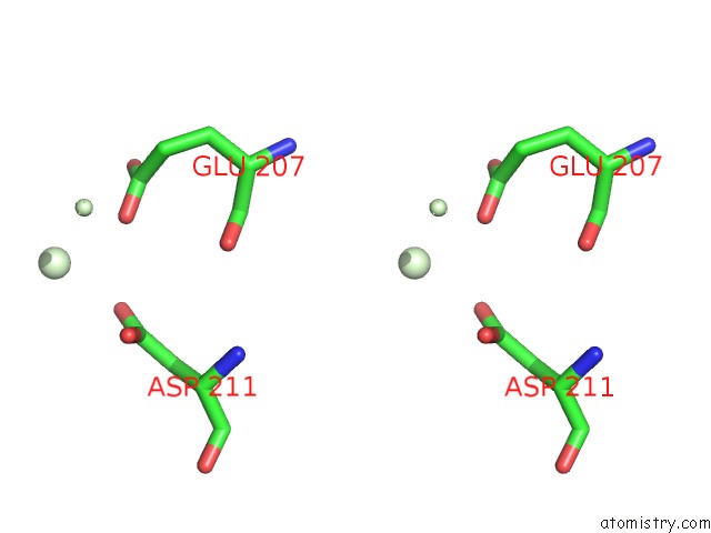
Stereo pair view
Mono view
Stereo pair view
A full contact list of Praseodymium with other atoms in the Pr binding
site number 1 of Protein B within 5.0Å range:
|
Praseodymium binding site 2 out of 2 in 6oop
Go back to
Praseodymium binding site 2 out
of 2 in the Protein B
Mono view
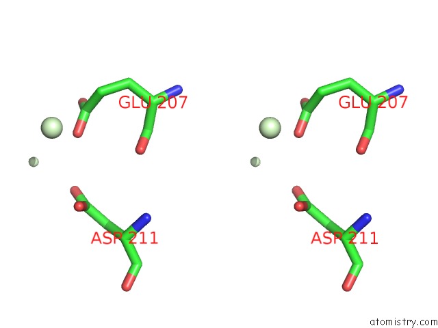
Stereo pair view
Mono view
Stereo pair view
A full contact list of Praseodymium with other atoms in the Pr binding
site number 2 of Protein B within 5.0Å range:
|
Reference:
H.H.Wu,
J.Symersky,
M.Lu.
Structure of An Engineered Multidrug Transporter Mdfa Reveals the Molecular Basis For Substrate Recognition. Commun Biol V. 2 210 2019.
ISSN: ESSN 2399-3642
PubMed: 31240248
DOI: 10.1038/S42003-019-0446-Y
Page generated: Thu Oct 10 10:31:17 2024
ISSN: ESSN 2399-3642
PubMed: 31240248
DOI: 10.1038/S42003-019-0446-Y
Last articles
Na in 2WGDNa in 2WGE
Na in 2WG4
Na in 2WG9
Na in 2WFT
Na in 2WFX
Na in 2WG8
Na in 2WG7
Na in 2WDV
Na in 2WF9