Praseodymium »
PDB 6e1a-9bif »
6vs2 »
Praseodymium in PDB 6vs2: Protein D
Protein crystallography data
The structure of Protein D, PDB code: 6vs2
was solved by
M.Lu,
M.M.Lu,
with X-Ray Crystallography technique. A brief refinement statistics is given in the table below:
| Resolution Low / High (Å) | 15.00 / 3.00 |
| Space group | C 1 2 1 |
| Cell size a, b, c (Å), α, β, γ (°) | 95.014, 66.973, 110.433, 90.00, 111.10, 90.00 |
| R / Rfree (%) | 28 / 29.9 |
Praseodymium Binding Sites:
The binding sites of Praseodymium atom in the Protein D
(pdb code 6vs2). This binding sites where shown within
5.0 Angstroms radius around Praseodymium atom.
In total only one binding site of Praseodymium was determined in the Protein D, PDB code: 6vs2:
In total only one binding site of Praseodymium was determined in the Protein D, PDB code: 6vs2:
Praseodymium binding site 1 out of 1 in 6vs2
Go back to
Praseodymium binding site 1 out
of 1 in the Protein D
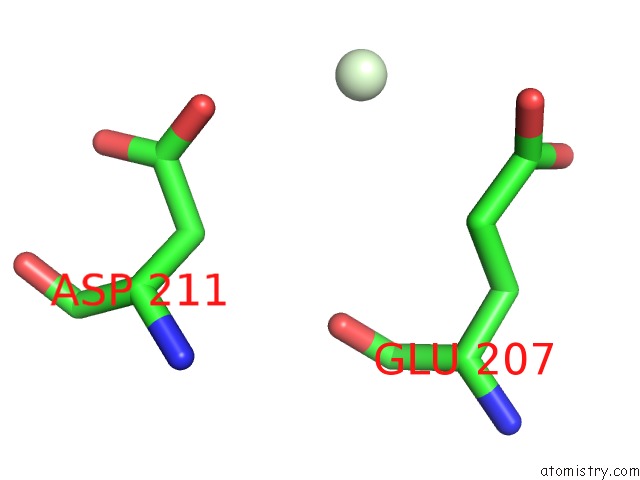
Mono view
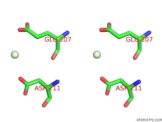
Stereo pair view
Mono view
Stereo pair view
A full contact list of Praseodymium with other atoms in the Pr binding
site number 1 of Protein D within 5.0Å range:
|
Reference:
H.H.Wu,
J.Symersky,
M.Lu.
Structure and Mechanism of A Redesigned Multidrug Transporter From the Major Facilitator Superfamily. Sci Rep V. 10 3949 2020.
ISSN: ESSN 2045-2322
PubMed: 32127561
DOI: 10.1038/S41598-020-60332-8
Page generated: Thu Oct 10 10:32:06 2024
ISSN: ESSN 2045-2322
PubMed: 32127561
DOI: 10.1038/S41598-020-60332-8
Last articles
Na in 5Z84Na in 5Z7W
Na in 5Z48
Na in 5Z42
Na in 5Z5O
Na in 5Z39
Na in 5YYD
Na in 5Z2V
Na in 5Z1N
Na in 5YZ3